1-2 Canadian Medical Association Journal, 2019
Could a Prostate MRI be Right for You?

DETECTION
Patients who have elevated PSA levels, as determined by their physician or urologist, may qualify for a prostate MRI.
Often, these patients may also have already had a negative biopsy, however, this is not necessarily required. In fact, recent studies* have shown that using MRI first provides more accurate information before proceeding to the biopsy. An MRI-first protocol is also associated with a reduced number of unnecessary biopsies.
Prostate MRI can also diagnose infection in the prostate, as well as benign prostatic hyperplasia (BPH) or congenital abnormalities.
Patients may also choose MRI for early detection through our Enhanced Prostate Screening program (EPS). These patients may be concerned about their prostate health, have a family history of prostate cancer, or simply want to take charge of their well-being.3

STAGING
For patients who have had a positive biopsy result and know they have prostate cancer, Prostate MRI is an advanced imaging technique that produces detailed pictures of the structures inside the prostate gland. These enhanced images allow us to precisely measure tumor size, extent and activity. We can also determine if the cancer is confined within the gland or has spread beyond the capsule. This information allows for accurate treatment planning, such as surgery or radiation therapy.

ACTIVE SURVEILLANCE & RECURRENCE
Patients who have previously been diagnosed with low-risk prostate cancers that are being managed with active surveillance, benefit from regular Prostate MRI’s to rule out occurrence of higher-grade elements.
Those who have previously been treated for prostate cancer with either surgery or radiation and have a PSA relapse will also benefit from Prostate MRI.
Prostate MRI is useful to help assess whether prostate cancer has returned.

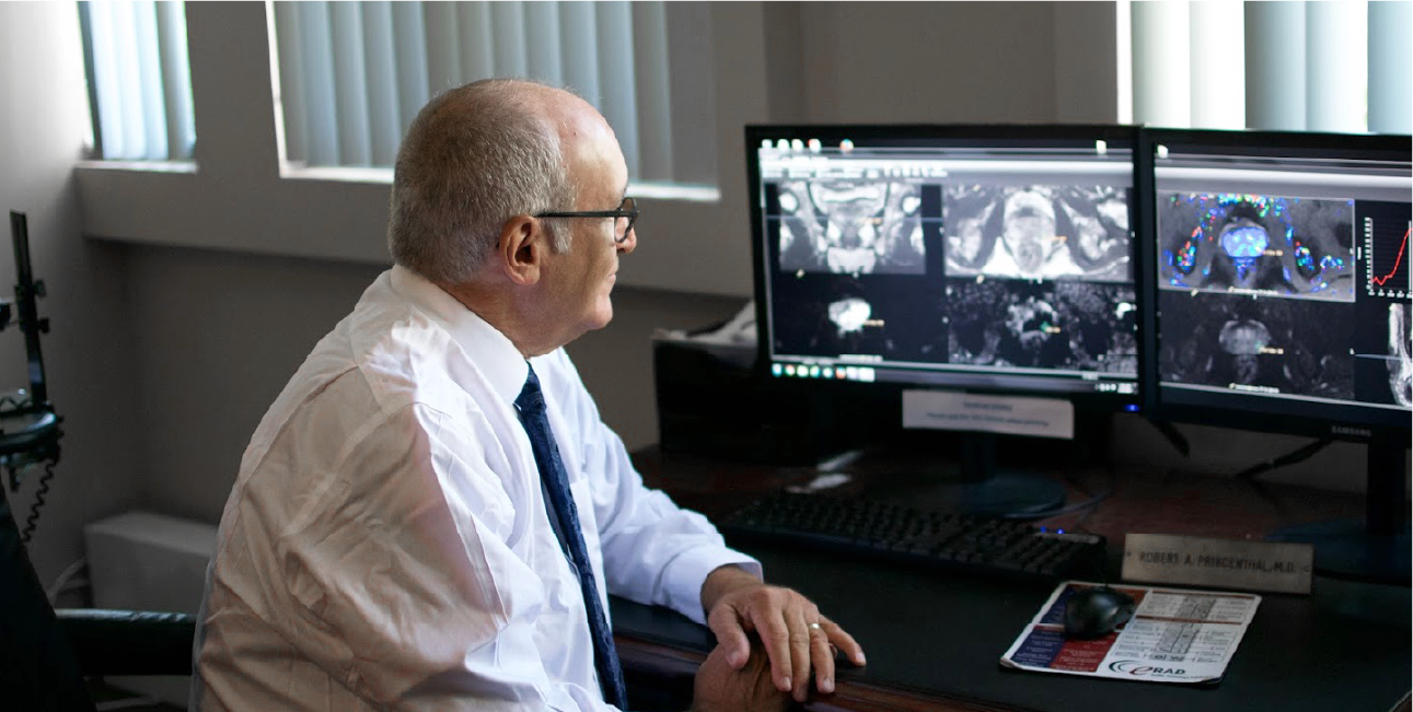
"The main value of obtaining a prostate MRI is that it can help distinguish which men would benefit from a biopsy to evaluate a rising PSA, versus which men likely don't have cancer and can avoid a biopsy."
Robert Princenthal M.D.
Radiologist, Prostate Medical Director
Multi-Parametric Prostate MRI
MRI (magnetic resonance imaging) is a medical imaging technique that allows doctors to view detailed information on tissue and blood patterns found in the human body. Through the utilization of high-powered magnets a MRI machine can cause tissues in the body, include theprostate, to assume different appearances, thus revealing abnormal tissue.
Our dedicated Prostate MRI Program uses a multi-parametric approach to prostate MRI imaging, which has shown increased sensitivity and specificity for cancer detection (around 85 – 90 percent).
- These improved results are based by critical analysis of four (4) specific imaging sequences.
- These include high anatomic quality T-2 weighted imaging and three functional sequences.
The functional sequences include:
- DWI (diffusion weighted imaging) analyzes the restriction of movement of water molecules seen with prostate cancer. DWI is quite accurate and correlates when positive to a specific Gleason Score upon a positive biopsy result.
- DCE (dynamic contrast enhancement), where a small amount of IV agent is injected to evaluate increased and abnormal bloodvessels seen with prostate cancer. These DCE images are obtained rapidly and analyzed with computer generated curves which look for increased flow and rapid washout.
- Images are obtained rapidly, 3 seconds per acquisition and reviewed to generate time function curves.
Benefits
Data from multiple early implementers of prostate MRI have explored the efficacy of prostate MRI and subsequent MRI-guided biopsy.
They found that the specificity and sensitivity for finding clinically significant prostate cancer (those with a Gleason score of 6 or higher and a volume exceeding 5 millimeters) was between 85 percent and 96 percent. Their data also show a negative predictive value around 95 percent, which means there was a 95 percent chance that no significant prostate tumor existed if prostate MRI did not identify a tumor.
If a tumor nodule was identified, it could be confirmed with a targeted, MRI-guided biopsy. The cancer detection rate for MRI directed biopsy is around 80 percent compared to 45 percent at time of first TRUS biopsy.
MRI is a More Accurate Way to Screen and Stage Prostate Cancer – Increasing Detection and Reducing Overdiagnosis (Based on Published Research):
- Prostate MRI before biopsy may help reduce unnecessary biopsies.1
- There are more than 1 million prostate biopsies being performed each year -- about 75 percent of biopsies are negative for cancer.2
- MRI may save some men with a high PSA from a biopsy and also helps identify the more aggressive types of prostate cancer that need immediate treatment.3
- MRI showed potential benefits in detecting clinically significant prostate cancer (PCa) without using prostate-specific antigen (levels) as its defining criteria.3
Studies Also Show that it Matters Where You Have Your Prostate MRI – Experience Counts!
- Experienced radiologists were more likely to detect clinically significant prostate cancer on MRI.5
- The accuracy of prostate MRI may differ based on factors including imaging technique… and reader experience.6

1 https://jamanetwork.com/journals/jamanetworkopen/fullarticle/2808559
2 https://www.emedicinehealth.com/what_percentage_of_prostate_biopsies_are_cancer/article_em.htm
3 https://www.health.harvard.edu/mens-health/mri-looking-better-for-detecting-prostate-cancer
4 https://www.ajmc.com/view/mri-screening-provides-advantages-for-detecting-clinically-significant-prostate-cancer
5 https://www.sciencedirect.com/science/article/pii/S2666168321000604
6 https://pmc.ncbi.nlm.nih.gov/articles/PMC8061889/
For Physicians
The RadNet Prostate MRI Program offers referring physicians the ability to enhance and improve their local TRUS biopsy program. We hope the prostate MRI information on this website is helpful to the medical community. The protocols established by the MRI Program can help to assist with a higher yield of diagnostic accuracy, reduce infections, and promote fewer complications. The Prostate Program works in conjunction with your practice to provide referring physicians more detailed information to make treatment choices for their patients.
The Program will send key color images to referring physicians to help guide medical providers through the report. Follow-up conversations are available with the reading radiologists and/or a Program co-director upon request. The Program’s protocols include:
- Detection Protocol: for patients with a negative TRUS biopsy and unexplained elevated PSA. The scan will consist of protocols T2 imaging, DCE and DWI. No endo coil, no spectroscopy. Routine MRI safety screen plus an IV contrast prep.
- Prostate Cancer Staging Protocol: for patients who have had a recent biopsy proving cancer. In addition to the detection protocol the scan will be performed with an endo coil for optimal resolution and MR spectroscopy and IV contrast for the DCE sequences. This protocol should be used for active surveillance patients.
- Bones and Nodes Protocol: if a patient has a high pretest probability of metastatic prostate cancer we will perform a Bones and Nodes Protocol. This includes imaging the pelvis, lumbar spine and thoracic spine with T1 weighted images and diffusion/STIR images looking for bone metastasis and abnormal lymph nodes.
- Recurrence Protocol: for patients with rising PSA following either RT or surgery.
We Want To Hear From You
Prostate MRI
Arizona: (602) 892-3640
Fresno: (559) 474-4292
Coachella Valley: (760) 843-4028
Victor Valley: (760) 999-1041
Inland Empire: (909) 450-0651
Antelope Valley: (661) 246-3682
Long Beach: (562) 299-6245
Los Angeles: (310) 309-6711
San Fernando Valley: (818) 480-7261
San Gabriel Valley: (626) 208-3520
Temecula: (951) 238-6043
Ventura: (805) 299-8835
Orange North: (657) 304-2330
Orange South: (714) 784-1689
Bakersfield: (661) 362-0142